imaging-lab logo

computational biomedical imaging research

fetal MRI segmentation, mesh rendering. doi:10.6084/m9.figshare.12950009

about

The imaging-lab facilitates high-performance computing, data management and reproducible research for computational biomedical imaging projects in the SickKids Research Institute supported by dedicated access to the Research‑IT High Performance Computing (RIT‑HPC) core facility.

Aims

  • provide systems for the collection, analysis and interpretation of imaging research data
  • enable scientific creativity and productivity
  • promote collaboration, open science and reproducible research

Funded by the Canadian Foundation for Innovation.

projects

MRI recon server

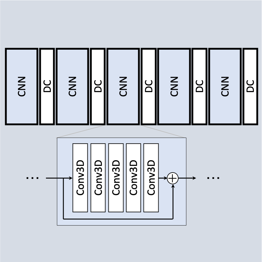
Integration and utilization of research MRI reconstruction methods can be hindered by computation lag and ease of use. An MRI reconstruction server was implemented using the Yarra framework, utilizing RIT‑HPC resources for computationally-intensive processes.

  • rapid prototyping of new methods
  • scalable resource allocation
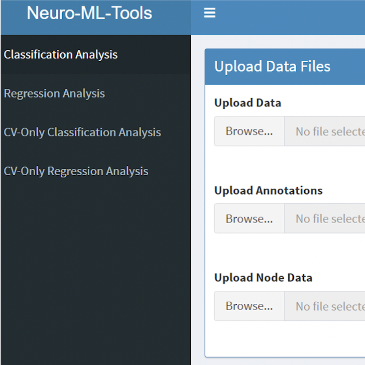
  • ease of use for clinically-focused researchers
  • automated end-to-end data transfer
  • systematic archiving of research data and output
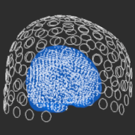
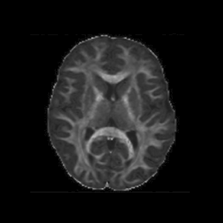
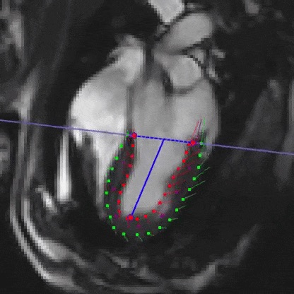
  • enables computation of motion-corrected fetal cardiac MRI

deep learning MRI recon

Reconstruction of under-sampled paediatric phase contrast MRI using a cascade of convolutional neural networks (CNN) with interleaved data consistency (DC). Image reconstruction with CNNs is fast, compared to conventional iterative compressed sensing algorithms, and better-suited to clinical deployment.
[Macgowan Lab]

  • developed in python using SigPy and PyTorch libraries
  • network trained using GPU-enabled RIT‑HPC node

MEGneto

Magnetoencephalography (MEG) processing and analysis pipeline implemented with MATLAB and the fieldtrip toolbox using imaging-lab’s computational power to analyze data from many participants.
[Mabbott Lab]

  • outputs functional connectivity matrices, time-frequency and spectral analyses
  • trial summaries, bootstrap significance testing, result visualization
  • interactive GUI to set analysis options, progress logging and email notifications
github.com/mabbottlab/megneto/

diffusion MRI

The Diffusion parameter EStImation with Gibbs and NoisE Removal (DESIGNER) pipeline was used to minimize thermal noise, Gibbs ringing and EPI spatial distortions, to improve the accuracy and reproducibility of diffusion processing. This diffusion MRI pipeline was implemented using MATLAB, MRtrix and FSL to process large cohorts.
[Mabbott Lab]

  • single python script that performs complete diffusion MRI processing
  • computational power facilitates fast execution time and handling of high resolution data

neuro-ml-tools

Toolkit and web app implementing supervised machine learning (ML) for downstream brain imaging data mining. The toolkit supports both regional features and functional/structure connectivity profiles from neuro data (MEG, fMRI, DTI and more), using recursive random forests, support vector machines and partial least squares regression/discriminant analysis.
[Dunkley Lab]

  • supervised feature selection
  • naïve data prediction
  • support for binary and multiclass classification, regression
  • report generation and results visualization
github.com/jzhangc/git_meg_ml_app/

AWSM-Q slide morphometry

Quantitative Automated Whole Slide Morphometry (AWSM-Q) segments images of histological samples and calculates morphological parameters to investigate rat models of lung disease.
[Santyr Lab]

  • Matlab-based histological analysis
  • enhanced processing capabilities allow for batches analyses
  • enables implementation of previously inaccessible methods

lung MRI quantification

Lung MRI analysis of 129Xe and 1H MRI images obtained from children with lung diseases for quantification of ventilation defect percentage.
[Santyr Lab]

  • Matlab-based co-registration and segmentation

cardiac strain analysis

Regional assessment of cardiac function to understand how the fetal heart repairs from myocardial infarction with the aim of finding the molecular pathways responsible for the proliferative response by the fetus and applying them in the adult heart. Calculation of radial, circumferential and longitudinal strain from MRI sheep experiments using cvi42.
[Seed Lab]

  • semi-automated segmentation of left and right ventricles
  • output as polar maps for regional assessment
  • 4D visualization

4D flow MRI

Visualization and analysis of complex blood flow patterns using cvi42 on workstations optimized for graphics performance.

containerization

Operating system virtualization to create and run 'containers' that package up software with dependencies using the Singularity platform. Containers can be built to include all of the programs, libraries, data and scripts such that comprise an entire experiment for reuse and sharing.

new users

New users should arrange the following accounts:

Contact the imaging-lab to discuss new projects.
Consult the user wiki for reference information and guidance.

Note: some linked pages only accessible via SickKids network

imaging-lab logo

SickKids Research Institute
  PGCRL 08.9470
imaging-lab.gitlab.io
imaging.lab@sickkids.ca